The current peer-reviewed literature on fluoroscopic evaluation of the pharynx and UES in dogs shows a lack of standardization regarding imaging protocol. The structure of the pharynx varies across the vertebrates.
Pharyngeal paralysis refers to paralysis of the upper throat pharynx that makes swallowing difficult or impossible.
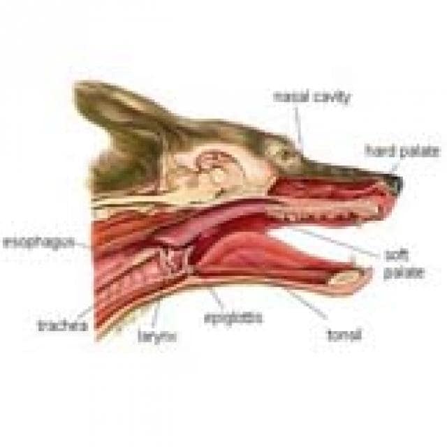
Where is the pharynx in the dog. The pharynx is the structure that lies at the back of the mouth and throat. It is the cavity behind the tongue and nasal passage through which both food and air are transported to deeper structures. The portion of the pharynx that is part of the respiratory tract is referred to as the nasopharynx and it connects the back of the nasal cavity to the larynx voice box.
In many other laboratory animal species eg dogs rats mice the pharynx is distal to most of the nasal airway and dorsal to the oral cavity and larynx. The pharynx is a musculo-membranous tube that is anatomically divided into nasal oral and laryngeal regions. 26-7 Radiograph of a dog evaluated for a cervical mass.
A mass located dorsal to the pharynx larynx and cervical portion of the trachea displaces these structures ventrally. There is effacement of the fascial planes in the cervical region. Carcinoma of salivary or sebaceous origin was diagnosed.
The pharynx comprises the caudal aspects of the oral and nasal cavities at the confluence of the digestive and respiratory tracts. The pharynx is a short fibromuscular tube divided into three compartments. The oropharynx the nasopharynx and the.
The structure of the pharynx varies across the vertebrates. It differs in dogs horses and ruminants. In dogs a single duct connects the nasopharynx to the nasal cavity.
The tonsils are a compact mass that points away from the lumen of the pharynx. Atlas of anatomy on x-ray images of the dog. This module of vet-Anatomy is a basic atlas of normal imaging anatomy of the dog on radiographs.
51 sampled x-ray images of healthy dogs performed by Susanne AEB Borofka PhD - dipl. ECVDI Utrecht Netherland were categorized topographically into seven chapters head vertebral column thoracic limb pelvic. Pharyngitis is inflammation of the walls of the throat pharynx.
It accompanies most upper airway viral and bacterial respiratory infections such as distemper in dogs. Other causes include damage of the pharynx by a foreign object or cancer of the mouth or tonsils. In dogs foreign objects stuck in the mouth and throat.
This condition is usually seen when a dog runs onto a stick being carried in the mouth. The stick impinges against the ground or another surface and the dogs weight drives the stick into the mouth and pharynx where it penetrateslacerates oropharyngeal tissues to varying degrees. May be acute dyspnea retchinggagging or subacutechronic.
Throat cone-shaped passageway leading from the oral and nasal cavities in the head to the esophagus and larynxThe pharynx chamber serves both respiratory and digestive functions. Thick fibres of muscle and connective tissue attach the pharynx to the base of the skull and surrounding structures. Both circular and longitudinal muscles occur in the walls of the.
The throat and pharynx in a dog. The upper throat is called the pharynx. Pharyngeal paralysis refers to paralysis of the upper throat pharynx that makes swallowing difficult or impossible.
The pharynx is the passage that is common to both the respiratory and digestive systems. It is located between the oral cavity proper and the esophagus. The pharynx is divided into three parts which you should identify.
Oropharynx nasopharynx and laryngopharynx. The base of the tongue toward the back of the mouth is bordered on the top by the nasopharynx which is the internal opening of the nostrils. The sides of the base of the tongue have folds which attaches it to the pharynx called the glossopharyngeal folds.
Read more dogs Pharyngitis in Dogs Pharyngitis is inflammation of the walls of the throat pharynx. It accompanies most upper airway viral and bacterial respiratory infections such as distemper in dogs. The radiographic anatomy of the pharynx and larynx of the dog is described based upon the examination of isolated specimens and radiographs of 140 clinically normal animals.
Variations within normality are described with particular reference to the mineralization of the laryngeal cartilages. The pharynx connects the nasal and oral airways with the laryngeal airway Figure 141. In humans and nonhuman primates the pharynx is situated posterior to the nasal cavity mouth and larynx.
In many other laboratory animal species eg dogs rats mice the pharynx is distal to most of the nasal airway and dorsal to the oral cavity and. Download scientific diagram Schematic representation of the pharynx in the dog with A normal B partial nasopharyngeal collapse and C complete nasopharyngeal collapse. Where Is the Pharynx.
The first thing to know about the pharynx is its location. The pharynx is below both the oral and the nasal cavities the mouth and the nose and just above the larynx and the esophagus. The pharynx extends all the way from the base of the human skull to the lower end of the cartilage known as the cricoid.
What is the pharynx and how does it relate to breathing. The pharynx is the area where the trachea and esophagus come together behind the tongue. The pharynx conducts air between the nose and the trachea and serves as the gatekeeper between the respiratory and gastrointestinal tract.
Measurement outcomes are significantly different between institutions and when bolus sizeconsistency is variable when assessing healthy dogs. The current peer-reviewed literature on fluoroscopic evaluation of the pharynx and UES in dogs shows a lack of standardization regarding imaging protocol. There is not a standard set of quantitative criteria.